Medovia Medical
Hospitals are under constant pressure to deliver timely imaging while patients become less mobile and clinical spaces become more crowded. The World Health Organization estimates that about 3.6 billion diagnostic X-ray examinations occur worldwide each year. That scale makes workflow, access, and equipment utilization important operational concerns. In intensive care units, emergency departments, operating rooms, and isolation areas, moving a patient to radiology may require several staff members, monitoring equipment, and valuable time. A portable X-ray machine brings the examination closer to the bedside. Sometimes, that is the safest option.
Why do hospitals prefer portable X-ray machines over fixed ones? The answer is rarely a single feature. Portable systems can support faster bedside imaging, reduce patient transfers, and help clinicians assess urgent conditions without disrupting treatment. They may also improve service continuity when a fixed room is occupied or unavailable. The FDA emphasizes that facilities must manage radiation exposure through justification, optimization, and appropriate training. The IAEA similarly highlights quality assurance, staff competence, and safe imaging practices as essential parts of radiology services. Technology alone is not enough.
This article explores seven practical reasons hospitals choose portable X-ray machines, from critical-care access and workflow flexibility to space efficiency and emergency preparedness. Real-world experience suggests clear advantages, but not every portable unit delivers identical image quality, battery life, or software performance. That is worth admitting. Procurement teams should compare clinical needs, detector performance, service support, cybersecurity, and total ownership costs before purchasing. A convenient device can still become an expensive mistake. Clear evidence and careful evaluation matter.
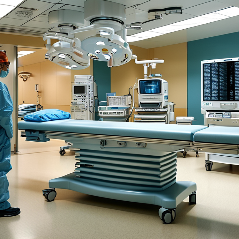
Portable X-ray machines are compact radiographic systems that travel to the patient, rather than moving the patient to a fixed room. A battery-powered generator sends electrons through an X-ray tube toward a metal target. The collision produces X-ray photons. These photons pass through the body and reach a digital detector, creating an image from differences in tissue absorption.
Hospitals choose them for seven practical reasons: bedside access, faster decisions, fewer patient transfers, emergency flexibility, intensive-care support, lower workflow disruption, and usefulness in crowded wards. A patient with breathing difficulty may remain connected to monitors while the machine rolls beside the bed. The image can then reach a radiologist through the hospital network. The IAEA and WHO estimate that billions of diagnostic X-ray examinations occur worldwide each year, showing why efficient imaging matters. Yet portable does not mean risk-free.
The UNSCEAR 2020/2021 report identifies medical exposure as the largest human-made source of ionizing radiation. Therefore, trained operators must confirm the indication, select exposure settings, limit the beam, and protect nearby staff. In practice, positioning is harder beside a bed. Image quality may suffer from movement, tubes, or limited detector space. That weakness deserves attention. A portable unit supports clinical judgment; it cannot replace careful examination, appropriate protocols, or qualified interpretation.
7 Tips Why Hospitals Choose Portable X-Ray Machines?
Why Hospitals Need Faster Imaging at the Point of Care
Portable X-ray machines bring imaging directly to the bedside. This matters when moving a patient could cause harm or delay treatment. Intensive care patients, trauma cases, and people on ventilators may need constant monitoring. A bedside scan avoids crowded corridors, elevator transfers, and interruptions to critical care. Less movement can also reduce staff workload and patient discomfort. Small details matter.
The Lancet Commission on Diagnostics estimated that 4.7 billion people lacked access to essential diagnostic services in 2021. Hospitals face a similar pressure locally: more patients, fewer imaging resources, and tighter response times. Portable systems help extend radiography into emergency rooms, intensive care units, recovery areas, and isolation spaces. Images can enter the digital workflow quickly, allowing clinicians to assess lines, lungs, bones, or post-procedure changes sooner. Faster acquisition does not guarantee faster decisions. Radiologist availability and network performance still matter.
A practical choice requires more than mobility. Hospitals should examine battery endurance, detector durability, infection-control procedures, image quality, and staff training. The World Health Organization emphasizes quality-assured diagnostic services, not simple equipment availability. A portable unit may be valuable, but poor positioning or rushed exposure can create repeat scans. That wastes time and increases workload. Clinicians should review repeat rates, report turnaround time, and patient-transfer incidents after implementation. The equipment is only one part of point-of-care imaging.
| Tip | Hospital Need | Portable X-Ray Advantage | Key Data Point | Typical Point-of-Care Use |
|---|---|---|---|---|
| 1. Faster bedside imaging | Clinicians need diagnostic information without waiting for transport to the radiology department. | The imaging system can be brought directly to the patient, helping reduce transport-related delays. | Exposure time is commonly measured in milliseconds | Emergency departments, intensive care units, recovery rooms, and inpatient wards |
| 2. Improved care for immobile patients | Moving critically ill, post-operative, ventilated, or trauma patients can increase clinical and logistical risk. | Bedside imaging avoids unnecessary transfers and helps keep monitoring and life-support equipment in place. | Useful when patient transfer is clinically unsuitable | Ventilated patients, intensive-care patients, fracture cases, and patients under isolation precautions |
| 3. Reduced workflow interruptions | Hospitals must coordinate transport staff, elevators, corridors, clinical escorts, and radiology-room availability. | Imaging can be performed in the treatment area, reducing dependence on transport resources. | One bedside examination can avoid multiple transfer steps | High-occupancy wards, isolation rooms, and periods of peak patient demand |
| 4. Flexible digital workflow | Radiology teams need images to be reviewed, stored, and shared efficiently. | Wireless or tethered digital detectors can send images to the hospital imaging network when configured for digital integration. | Common detector size: approximately 35 × 43 cm | Chest, abdominal, skeletal, and post-procedure radiography at the bedside |
| 5. Support for infection-control procedures | Hospitals need to limit unnecessary movement of patients with transmissible infections. | A portable unit can be used in or near the patient’s room, subject to local cleaning and radiation-safety protocols. | Cleaning is required between patient encounters | Isolation rooms, respiratory-care areas, infectious-disease units, and outbreak response |
| 6. Greater departmental capacity | Fixed radiography rooms may become bottlenecks when demand rises or patient acuity is high. | Portable systems provide an additional imaging pathway and can help distribute workload across care areas. | Can be deployed across multiple hospital zones | Overflow areas, temporary wards, disaster response, and after-hours coverage |
| 7. Better access in challenging locations | Some patients need imaging where a fixed X-ray room is unavailable or difficult to access. | Battery-powered, mobile designs can support imaging in treatment bays and other areas when safety requirements are met. | Battery operation supports operation away from a fixed room | Pre-hospital receiving areas, operating-room support, rehabilitation units, and remote clinical settings |
How Portable X-Ray Machines Improve Patient Safety and Comfort
Tip 1: Keep fragile patients in bed. Portable imaging avoids transfers that can cause pain, falls, or line displacement. AHRQ reports that approximately 700,000 to 1 million hospitalized patients fall each year in the United States.
Tip 2: Reduce corridor exposure. Bedside imaging limits movement through busy clinical areas. This may support infection-control practices, although portable X-ray use alone cannot prevent infections. The CDC estimates that about one in 31 hospital patients has a healthcare-associated infection on any given day.
Tip 3: Support faster clinical decisions. A radiographer can image a patient in the intensive care unit, emergency department, or recovery room. Less waiting can reduce anxiety and help clinicians respond sooner.
Tip 4: Improve physical comfort. Patients with fractures, breathing difficulty, or surgical wounds avoid unnecessary repositioning. Small adjustments still matter.
Tip 5: Protect vulnerable patients. Bedside imaging can help people who depend on ventilators, monitors, or multiple infusions. Staff should confirm equipment clearance before moving the detector.
Tip 6: Apply radiation controls carefully. The IAEA recommends justification, optimization, and dose limitation for occupational exposure. Portable examinations require barriers, distance, and trained operators.
Tip 7: Check image quality, not convenience alone. A repeat exposure increases discomfort and radiation dose. Good positioning remains essential, even beside the bed.
The World Health Organization’s Global Patient Safety Report 2024 states that about one in ten patients experiences harm in healthcare, with more than half considered preventable. Portable imaging cannot solve every risk. It can, however, remove one avoidable journey from a patient’s care. That benefit depends on competent staff, clear protocols, equipment checks, and honest review of failed examinations.
Portable X-ray machines bring imaging to patients who cannot safely travel. They support diagnosis in intensive care, emergency rooms, operating theatres, neonatal units, and isolation areas. The World Health Organization reports that one in ten patients experiences harm during healthcare, with over half considered preventable. Faster bedside imaging may reduce transfer risks, but it cannot replace clinical judgment.
Tip 1: Use portable imaging for unstable patients. A bedside chest image can check line placement, lung changes, or suspected fractures.
Tip 2: Prioritize intensive care workflows. Radiographers should confirm patient identity, positioning, and exposure settings.
Tip 3: Protect staff and patients. Apply local radiation-safety rules, distance controls, and shielding when appropriate.
Tip 4: Build quality checks into every shift. The International Atomic Energy Agency recommends structured quality assurance for diagnostic radiology equipment. Small faults matter.
Tip 5: Connect images with the electronic health record quickly.
Tip 6: Train teams for crowded bedside conditions. Cables, oxygen tubes, and urgent movement can affect positioning.
Tip 7: Review repeat-image rates. A repeat may reflect motion, poor centering, or an unsuitable technique.
Mobility is not magic.
A portable unit can produce a weak image when workflow becomes rushed. The WHO Global Patient Safety Action Plan 2021–2030 emphasizes safer systems, communication, and learning from errors. Hospitals should therefore measure image quality, reporting time, repeat exposures, and clinician satisfaction. These details show where portable X-ray machines genuinely support hospital diagnosis.
Hospitals usually examine seven factors before choosing a portable X-ray machine: image quality, radiation dose, battery life, weight, mobility, workflow, and service support. Image quality must remain reliable in crowded wards, intensive care units, and emergency rooms. The IAEA reports that medical imaging involves billions of examinations worldwide each year, making dose optimization a daily responsibility. Ask for documented performance data, not attractive demonstrations.
Dose matters. A suitable system should support justification, optimization, and clear exposure controls under IAEA safety guidance. Battery endurance affects how many bedside examinations staff can complete during one shift. A lighter unit reduces strain when elevators are busy or corridors become blocked. Small details matter. Hospitals should also check detector durability, wireless transfer, cleaning compatibility, and cybersecurity controls. The FDA’s guidance on medical X-ray imaging emphasizes safety, performance, and quality assurance throughout the equipment’s use.
Workflow can decide the purchase. Test whether images reach the hospital archive quickly, even during peak hours. Review maintenance response times, training, warranty terms, and replacement-part availability. Total cost includes software updates, battery replacement, servicing, and staff downtime. The choice is not always obvious. A technically advanced machine may perform poorly if nurses find it difficult to position or clean. In practice, hands-on trials with radiographers and nurses reveal problems that brochures often miss. Industry reports can guide evaluation, but local workflow evidence should carry greater weight.
: It is a compact imaging system that travels to the patient’s bedside. It can operate in wards, emergency rooms, and intensive-care areas. It brings imaging closer.
A battery-powered generator sends electrons through an X-ray tube. The electrons strike a metal target and produce X-ray photons. The photons pass through the body and reach a digital detector.
They reduce patient transfers and support faster clinical decisions. A patient can remain connected to monitors while the machine rolls beside the bed. This matters in crowded wards.
No. Ionizing radiation still requires careful control. Qualified operators should confirm the need, limit the beam, and select suitable settings. Nearby staff may also need protection.
Movement, medical tubes, and limited detector space can affect the image. Positioning beside a bed is often difficult. That weakness deserves attention.
Hospitals should assess image quality, radiation controls, battery life, weight, and mobility. They should also check detector durability, cleaning compatibility, wireless transfer, and cybersecurity controls. Small details matter.
Battery endurance affects the number of examinations completed during one shift. A lighter unit is easier to move through blocked corridors and busy elevators. That can reduce staff strain.
Staff should test image transfer during peak hours. Radiographers and nurses should practice positioning, cleaning, and daily movement. A brochure can look convincing. It can still mislead.
Total cost may include software updates, battery replacement, servicing, training, and staff downtime. Maintenance response and replacement-part availability also matter. The cheapest purchase may not be the cheapest solution.
No. It supports clinical judgment but cannot replace examination or qualified image interpretation. Appropriate protocols remain necessary. The choice is rarely obvious.
Portable X-ray machines are compact imaging systems designed to be moved directly to a patient’s bedside. They use controlled X-ray exposure and digital detectors to produce diagnostic images without requiring the patient to travel to a radiology department. Why do hospitals prefer portable X-ray machines over fixed ones? The main reasons are speed, flexibility, and improved point-of-care service. They help medical teams obtain images quickly in emergency rooms, intensive care units, operating rooms, recovery areas, and isolation wards.
By reducing patient transfers, these systems can improve comfort, lower the risk of falls or equipment-related complications, and support safer care for critically ill or immobile patients. Hospitals also consider image quality, battery performance, ease of operation, radiation protection, maneuverability, maintenance needs, and total cost before making a selection. Overall, portable X-ray machines help healthcare professionals make faster diagnostic decisions while providing convenient and patient-centered imaging across different hospital environments.